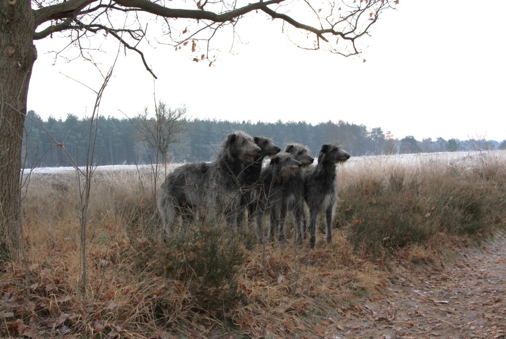
 Thrombotic Disease in Deerhounds—A “New” Blood Clotting Disorder
Emerging information in Greyhounds suggests that Deerhounds may experience a third type of clotting disorder called thrombotic disease, in which a blood clot forms in the wrong place or at the wrong time.
by John Dillberger, DVM, PhD
Reprinted from the November/December 2020 issue of The Claymore.
I have written in the past about two blood clotting disorders in Deerhounds that cause prolonged or inappropriate bleeding: Factor VII Deficiency and Hyperfibrinolysis Syndrome, also known as Delayed Postoperative Bleeding Syndrome. Both disorders occur in other breeds.
Factor VII Deficiency can slow clot formation, so that a dog bleeds excessively from a wound, either accidental or surgical. The disease occurs when a dog inherits two copies of a variant of the Factor VII gene.
Hyperfibrinolysis Syndrome probably also is inherited, but the genetic basis is not yet worked out. Fibrinolysis (pronounced “fib-rin-ALL-iss-iss”) is the medical term for the process by which a blood clot dissolves after a damaged blood vessel has healed. It comes from the words “fibrin” (rhymes with hyphen) which is the sticky, threadlike protein that makes up a clot, and “lysis,” which is Greek for freeing or unbinding. Dogs with Hyperfibrinolysis Syndrome make blood clots well, but the clots dissolve too fast. Affected dogs do not bleed excessively during surgery. Instead, they begin to bleed at the surgical site one or two days after surgery.
Emerging information in Greyhounds suggests that Deerhounds may experience a third type of clotting disorder called thrombotic disease, in which a blood clot forms in the wrong place or at the wrong time. The name comes from the medical term for a blood clot (“thrombus”). The process of clot formation is called “thrombosis.”
The consequences of thrombotic disease can be just as dangerous as those from a bleeding disorder. A blood clot in the wrong place reduces or cuts off blood flow to downstream tissues, which can die from lack of oxygen. If the clot blocks a large blood vessel, then it can raise blood pressure behind the clot, putting a strain on the heart.
Over the past 30 years, I have learned of many Deerhounds that developed blood clots in the wrong place. Sometimes this was discovered only during surgery or at necropsy. The same is true in humans, where thrombosis often is challenging to diagnose, even when suspected.
For example, readers may recall my 2015 column about Lynn Robb’s 7-year-old male named Ioan. Ioan had multiple health issues, one of which was a blood clot in the vein leaving his spleen that was found during an ultrasound examination. At necropsy, the pathologist discovered Ioan also had a blood clot in his portal vein, which carries blood from the intestinal tract to the liver. The 2011 Deerhound Health Survey includes entries for a 4 1⁄2 year-old bitch that, like Ioan, had thrombosis of the splenic and portal veins, and a 4-year-old male that had “suspected liver and gastrointestinal tract thrombosis” based on surgical and necropsy findings. My own 4-year-old bitch experienced sudden, fatal, pulmonary thrombosis while we were at the National Specialty in Harrisburg, Pennsylvania.
Recently, Barb Heidenreich pointed me to a new paper by Dr. Guillermo Couto that included a review of thrombotic disease in Greyhounds. If you are a regular reader of my column, you may recognize Dr. Couto as the veterinary researcher who identified and studied Hyperfibrinolysis Syndrome in Greyhounds and who carried out clinical trials of a drug (aminocaproic acid or Amicar®) that could prevent and treat the disease. The treatment that he pioneered has prevented this costly and potentially fatal disorder in Deerhounds as well as Greyhounds.
This month, I want to share information about thrombotic disease in Greyhounds. Based on my experience and on similarities in the physiology of Greyhounds and Deerhounds, I think it is likely that thrombotic disease also occurs in our breed. If so, then just as with Hyperfibrinolysis Syndrome, knowledge gained from Greyhounds may benefit Deerhounds.
Thrombotic Disease in General
Two medical terms come up repeatedly when discussing thrombotic disease, both coined by pathologist Rudolph Virchow in the 1850s. One is “thrombus,” which is another word for a clot. The other is “embolus,” which is the Greek word for a wedge-shaped object. And embolus is an abnormal particle floating in the blood, such as an air bubble, piece of fibrocartilage, or piece of a thrombus. Embolism is dangerous because the embolus can lodge in and block a blood vessel. When the embolus is a blood clot fragment, the situation is called “thromboembolism,” often abbreviated as TE. TE is a potential consequence of any thrombotic disease.
Thrombotic disease can be caused by local factors. For instance, damage to the blood vessel lining will cause a clot to form at the damage site. This is what causes most heart attacks in humans, where atherosclerosis in a coronary artery causes a clot to form that restricts or totally blocks the blood supply to part of the heart. Another local factor that can cause a blood clot to form is slowing of blood flow. This is what happens in dogs and humans that have a heart arrhythmia called atrial fibrillation. A fibrillating (vibrating) atrium cannot pump blood, and the stagnant blood inside the atrium is prone to clotting.
Thrombotic disease also can be caused by so-called systemic factors that sensitize the blood clotting system, making it prone to form clots. For example, when a dog is injured, particularly if the injury is substantial, one of the many responses is for the blood clotting system to become sensitized to form clots. From an evolutionary perspective, this offers a clear advantage, as it reduces the chance of life-threatening blood loss from injuries that happen when hunting or fighting.
Another situation that increases the risk of thrombotic disease is an abnormally high number of red blood cells in circulation. This not only makes the blood thicker, but it also makes the blood more prone to form clots. The underlying reason is unknown, although one possibility is that it simply increases collisions between blood cells and blood vessel walls, leading to more blood vessel damage.
Thrombotic Disease in Greyhounds
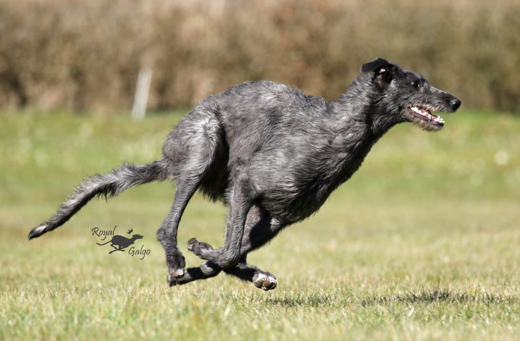
Greyhounds and Deerhounds have more red blood cells circulating, on average, than do non-sighthound breeds, which could make Greyhounds and Deerhounds more prone to thrombotic disease. On the other hand, both breeds also have fewer platelets and a greater antithrombin activity (an enzyme that inhibits blood clotting) in their blood, which may counterbalance that risk.
Exercise and excitement can increase the number of red blood cells in circulation. This has been studied in some detail in Greyhounds. Shortly after starting a race, a Greyhound has a surge of adrenaline. Among other things, this causes a massive contraction of the spleen, which is a reservoir of both red blood cells and platelets. When the spleen contracts, the number of circulating red blood cells and platelets suddenly increases. In some Greyhounds during or immediately after a race, the blood can be 80% cells, rather than the usual 55%! This increase in red blood cells clearly improves performance by delivering oxygen more efficiently to the dog’s muscles, and it also increases the blood’s ability to neutralize the lactic acid being produced by those muscles. But it also makes the blood more viscous and makes the heart work harder to move the blood around. And it also could increase the risk of clot formation.
Several sorts of thrombotic disease have been reported in Greyhounds. For instance, there are reports of TE in the brain or spinal cord of Greyhounds and other sighthounds. Recent reviews also described thrombosis in the aorta and iliac arteries of several Greyhounds and TE in the lungs of a Greyhound. Although some of the non-Greyhound dogs in these reports had underlying disorders, such as cancer or heart disease, most of the Greyhounds and other sighthounds affected were otherwise healthy.
In most Greyhound patients managed by Dr. Couto or described by others, underlying coagulation abnormalities were not identified using an extensive battery of lab tests. He notes that old reports (1970s) suggest that Greyhounds and other sighthounds may have tortuous arteries, develop hardening of the arteries (arteriosclerosis), or develop fatty deposits in artery walls (atheromas). Any of these could explain the higher prevalence of thrombotic disease in sighthounds. However, there are no recent reports of any of these underlying disorders in a sighthound breed.
Thrombotic Disease in the Brain (Stroke)
In Dr. Couto’s experience, TE in the brain is common in Greyhounds and other sighthounds. TE in the brain is like a stroke in humans. Here is how Dr. Couto explains stroke in Greyhounds, with some of the medical terms translated:
“The typical presentation is that of an older (8- to 10-year-old) dog, with peracute [sudden] onset of CNS [central nervous system] signs consisting of ataxia [unsteady gait], salivation, nystagmus [rapid, rhythmic eye movement], seizures, or any combination of neurologic signs; in my experience, most dogs present with central vestibular signs [loss of balance, head tilt, circling tendency]. Results of magnetic resonance imaging (MRI) typically reveal a well-defined lesion that… involves predominantly the gray matter in the vascular territory of main cerebral or cerebellar arteries or a perforating branch of such arteries. Most dogs have a single lesion.”
A 2006 study of 40 dogs with stroke (Garosi et al.) found that Greyhounds were the second most commonly affected breed (13%), surpassed only by Cavalier King Charles Spaniels (20%). A 2014 study of 21 Greyhounds with strokes (Kent et al.) found that the age at which stroke occurred ranged from 2 to 12 years. Most affected Greyhounds evaluated by Dr. Couto or reported by Kent et al. had normal blood coagulation test results.
Thrombotic Disease in the Spinal Cord (Spinal Stroke)
Dr. Couto also has identified a subset of young, actively racing Greyhounds that develop peracute spinal cord signs within 50 to 75 meters of starting the race. As he explains:
“Most trainers and veterinarians refer to [the cause] as fibrocartilaginous emboli (FCE), despite the fact that, to my knowledge, no imaging or pathologic studies have documented the lesions. Although we have limited data from advanced imaging, most affected dogs that underwent MRI had evidence of arterial thrombosis. In most dogs, the response to antiplatelet agents is rapid (hours), suggesting a thrombus as opposed to FCE. Interestingly, these episodes seem to coincide with the purported splenic contraction secondary to cate- cholamine release that rapidly increases circulating red blood cell count. In addition to the arterial thrombosis mentioned above, we have seen peracute thrombosis of the spinal venous sinuses in pet Greyhounds as young as 18 months of age.”
This paragraph caught my eye. You may remember that the January/February 2013 issue of The Claymore contained an article by Harriet King entitled “Bryce’s Tale: A Saga of FCE.” Harriet’s young Deerhounds, JD and Bryce, had their usual morning play session, and then each went to his crate for a nap. When Harriet checked an hour later, Bryce was paralyzed from the neck down. She immediately took him to a veterinary neurologist at the Veterinary Specialty Hospital at UC Davis, where he was diagnosed with FCE. He was given the standard treatment—corticosteroids and antibiotics—followed by weeks of physical therapy.
I personally witnessed an episode of apparent FCE in a Deerhound many years ago at a National Specialty in Vergennes, Vermont. Betty Stephenson and I both were called over to examine a dog that had suddenly become weak and wobbly in his hind quarters. Examination revealed deficits in reflexes in the hind limbs. It was clear that the dog had some acute episode of damage to his lumbar spinal cord. I do not remember who the owner was or what became of the dog.
Treating Stroke
Dr. Couto manages dogs with stroke in the brain or spinal cord using anti-platelet drugs. Aspirin is his first choice. In his own words:
“In my experience, aspirin at a dosage of 40.5 mg (half of an 81-mg aspirin) per dog by mouth, every 24 hours], is effective in most dogs treated shortly after the development of clinical signs. Indeed, in some dogs, administration of aspirin at home by their owners frequently results in marked improvement or even resolution of most neurologic signs by the time the patient arrives at the clinic. We continue this treatment indefinitely, and thus far, I have documented only two dogs who relapsed.
“…we documented that 40.5 mg of aspirin given [by mouth] once daily significantly decreases platelet aggregation in Greyhounds. We were unable to demonstrate decreases in platelet aggregation in a small number of Greyhounds in which we used clopidogrel (Plavix); this may be due to the fact the latter is a cytochrome P450 (CYP450)-dependent drug. Anecdotally, however, clopidogrel may be beneficial in Greyhounds.” (S. Shropshire, DVM, DACVIM, Colorado State University, personal communication).
Thrombotic Disease in the Aorta in Greyhounds
Greyhounds can develop a blood clot in their aorta, often at the end where it splits into two iliac arteries that supply blood to the hind legs. Affected Greyhounds usually have intermittent, progressive, rear limb lameness that frequently starts in one leg and gradually affects the other. Swelling and bruising of the affected limb are common. In most affected Greyhounds, physical examination also reveals a difference in the strength of the femoral pulses between the two legs. Ultrasonography is the best imaging method of diagnosis.
A 2012 paper of 31 cases of aortic thrombosis (Lake-Bakaar et al.) stated that no breed was overrepresented in the case series. But in a recent study of 100 dogs with aortic thrombosis (Ruehl et al., 2020), Greyhounds were the second most common breed, accounting for 14% of the cases. They were almost tied with the most common breed (Labrador retrievers), which accounted for 16% of the cases.
Treatment for thrombotic disease in the aorta is aspirin, as described for stroke. In a Greyhound that does not respond to aspirin therapy or that has severe edema/ bruising, Dr. Couto has successfully used warfarin; however, this is best done under the supervision of a veterinary specialist, as the dosage needs to be adjusted based on coagulation analyses.
The picture that has emerged is that Greyhounds probably have a greater risk of thrombotic disease than dogs in general. To the extent that the increased risk is due to a physiological factor, such as higher red blood cell counts, then the risk probably is shared with other sighthound breeds, including Deerhounds. My own anecdotal experience suggests so.
From a practical perspective, there are two consequences for Deerhound owners. First, it is important to add thrombotic disease to the list of possible causes for illness in our breed. And I think that thrombotic disease should be at the top of the list for a Deerhound that experiences sudden symptoms of brain damage, such as stroke-like signs, head tilt, or circling, or sudden symptoms of cord damage, such as weakness or paralysis in one or more legs. Likewise, in a Deerhound with symptoms of liver disease or splenic disease, thrombosis should be one possibility investigated.
The second practical consequence is that most veterinary practitioners will not be familiar with the emerging information about thrombotic disease in Greyhounds, and even fewer practitioners will think of applying that information to Deerhounds. For that reason, it might be a good idea to print this article and give it to your veterinarian, just to put the issue on her or his radar screen.

References
Couto GC (2020). “Why do some Greyhounds bleed and others clot excessively?” Veterinary Practice News at https://www.veterinarypracticenews.ca/why-do-some-Greyhounds-bleed-and-others-clot-excessively/.
Garosi L, McConnell JF, Platt SR, Barone G, Baron JC, de Lahunta A, Schatzberg S.J (2006). Clinical and topographic magnetic resonance characteristics of suspected brain infarction in 40 dogs. Journal of Veterinary Internal Medicine 20: 311–321.
Kent M, Glass EN, Haley AC, March P, Rozanski EA, Galban EM, Bertalan A, Platt SR (2014). Ischemic stroke in Greyhounds: 21 cases (2007–2013). Journal of the American Veterinary Medical Association 245: 113–117.
Lake-Bakaar GA, Johnson EG, Griffiths LG (2012). Aortic thrombosis in dogs: 31 cases (2000–2010). Journal of the American Veterinary Medical Association 241: 910-915.
Ruehl M, Lynch A, O’Toole TE, Morris B, Rush J, Couto C, Hmelo A, Sonnenschein S, Butler A, Guillaumin J (2020). Prognostic indicators in dogs with aortic thrombosis: 100 cases (1997-2014). Journal of Veterinary Internal Medicine at https://onlinelibrary.wiley.com/doi/epdf/10.1111/jvim.15874.